|
The
Digestive System and Urinary Tracts |
On this page you can read all about the
digestive system and the urinary tracts
so that the
information on the pages that follow will be
a little easier to understand.
*
The Digestive System
*
Urinary Tracts

Picture source:
Gezondheidsnieuws
The digestive System
The intestines actually determine what may and may not get in our body. They have a filter function and thus protect us against all infections and inflammations. A whopping 80% of the immune system is affected by the action of the bowel. If something goes wrong in the bowel, then there are consequences in the body almost everywhere. The intestines have a direct impact on the lungs, skin, liver, gall bladder, stomach, pancreas, spleen, nasal cavities and the heart. If waste is not properly discharged by the intestines these substances pile up or seek a way out through the skin causing pimples. As many as 1 in 7 Dutch, about 2 million people, regularly suffer from gastro-intestinal complaints. Nearly half of them have chronic problems. There may be some 200 different disorders of the stomach, liver, intestinal tract which can occur. For example, some 50,000 Dutch people suffer from chronic bowel inflammation, including Crohn's disease and ulcerative colitis. The following factors can cause intestinal symptoms: unusual food, stress, aging, virus/infection/disease, parasites, use of antibiotics (which kills the good as well as the bad bacteria), medication/radiation/surgery, insufficient movement, alcohol, smoking and the climate.
Source: Zin gezond

Picture source:
Erfelijkheid.nl
The entire digestive tract (esophagus, stomach, liver, pancreas, intestine and rectum) has a length of about 9 meters. Every day about 11 liters of food, liquids and digestive juices run from the digestive tract. Our digestive system is unique: it can digest meat, vegetables and cereals. It normally takes a meal 24 - 30 hours to go through completely. It spends the most time in the large intestine and the rectum. Ultimately about 1.5 liters of water, along with the indigestible food, goes into the large intestine. The stools contain just a deciliter (100mls) of water. An average adult has 100 - 150 grams a day of stools. During a lifetime the average gut has 65,000 pounds of food and drink to process (the weight of 12 elephants!).

It begins in the mouth (cavum oris) where the food is chewed and mixed with saliva. In the saliva are enzymes, which help digest food. Salivary glands produce saliva not only as if food enters the mouth, but also if you see or smell food, and even if you think about it. By day you produce about 1200 ml of saliva. The composition depends on the type of food. For a good digestion you should chew each bite 40 times. After swallowing, the nasal cavity is closed by the uvula and the trachea by the throat cover, the food goes via the throat (pharynx) into the gullet (esophagus), which is a 25 cm long flexible tube with a diameter of 2.5 cm. From here the body regulates the digestion itself: you do not consciously do it. The food very quickly goes through the esophagus, on its way to the stomach (ventricles, gaster). Because of the peristaltic movements of the esophagus, you can still eat and drink if you lie down, or even if you are standing on your head.

Picture source:
cancerresearch
During the transition between the esophagus and stomach, a round circular muscle (cardia sphincter) ensures that no food and stomach acid flows back into the esophagus. When there is food in the mouth, the stomach gets a message from the brain know that food is on the way and immediately of the stomach begins to prepare the creation of acids. The stomach is a muscular bag with folds (rugea) which serve as food reservoirs and can contain 3 to 4 liters of water and food. Without food the stomach is completely flat, and when eating a kind of reversed pear. The stomach consists of 2 parts: in the upper part (the cardia part) the acid and pepsin production takes place (pepsin is a protein splitting enzyme. And in the lower part (atrium) of the stomach certain cells form the hormone gastrine, which stimulates production of gastric juices. The stomach breaks the food up by continuously mixing and kneading it with gastric juices, until it is a thick paste (chymus). The stomach takes about 3 to 4 hours (depends on how fatty the food is) for a meal to be fully digested. At the bottom of the stomach is a door (pylorus),which allows small quantities of liquid to pass the 25 cm long twelve fingered colon (as the first part of the small intestine (interstinum tenue ) is called, also knows as the duodenum). The food is already crushed into particles of about 1 millimeter. The drainage of the pancreas and gall bladder also comes out in the duodenum. That happened on 1 common opening: the papilla of Vater.

Picture source:
gerd.com
Nutrients are absorbed in the small
intestine. As the intestine makes kneading movements the
nutrients can already be absorbed by the blood through
the intestine wall. The peristaltic movements push the
chime through the small intestine at a speed of more
than one centimetre per second in approximately 4.5
hours. At the end of the small intestine, a length of
some 6 to 7 metres, almost all the useful nutrients have
been absorbed.

The inside of the small intestine is covered with what
are known as villi, sort of tiny projections which
ensure that the surface where the nutrients can be
absorbed is considerably increased.
Picture source: from the book "Atlas van
de anatomie".
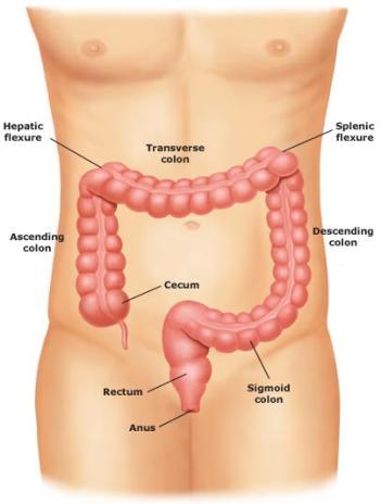
The rest of the food than goes to the 1.5 meter long large intestine (cartel intestine, or colon interstinum crassum), looking like a kind of inverted "U" which is located in the abdominal cavity. The colon is distinguishable from the small intestine because it is much wider, with at regular intervals pockets (haustra) with hollows in between (plicae semilunares; embryo created by fold formations in the wall) to be seen and three longitudinal muscle bands. The colon runs from the appendix (caecum) in the lower right abdomen, first straight up (colon ascendant / ascending colon) to the liver. Then the colon turns sharply to the left (colon transversum / transverse colon). Under the spleen it bends back down (descendent colon / descending colon). Then the colon makes an s bend forward (colon sigmoideum or simply the sigmoid) and afterwards turns into the rectum (rectum).
In the appendix are 2 large mucosal folds (Bauhini doors), to prevent food flowing back to the small intestine. At the bottom of the appendix is a worm-shaped Appendix: the appendix vermiformis. When one speaks of "appendicitis", it is really this inflamed appendix.
Picture source:
Uptodate.com
Just like the small intestine, the wall of the large
intestine consists of 3 layers. From the outside inwards:
the double layer of muscle, the layer of connective
tissue and the layer of mucous membrane. The mucous
membrane of the large intestine certainly has folds but
there are no villi. The total surface area of the large
intestine mucous membrane is much smaller (4m²) than
that of the small intestine (150-200m²).
Picture source:
Academie voor Mesologie
The most important function of the large intestine is to
extract the fluid and salts from the excrement and
to
reabsorb them into the body. As a result the excrement
becomes thicker and is changed into a form that the body
can eliminate easily. The large intestine produces
mucous that acts as a lubricant. In the large intestine
there are enormous numbers of bacteria, around 100
billion and more than 400 types. These are known as
intestinal flora (see illustration here below).
Intestinal flora consist of useful and harmful bacteria.
In healthy flora these bacteria are in balance with one
another. Intestinal flora are responsible for the
fermentation
and breakdown of the content of the
intestine, whereby matter is released which stimulates
the movements of the large intestine. Healthy intestinal
flora also protect us from infections. If the harmful
bacteria gain the upper hand then we can fall ill.

Picture source:
Quarks & Co
The large intestine (colon) is characterized by its haustra. These are folds of the intestine along the outside every few centimeters seems like cords. The haustrale contractions are slow movements and last 1 minute, and happen every 30 minutes. On the images below you can see how the stool (Latin: faeces) in the colon is formed. Mixing of the large-intestinal contents happens due to local contractions of the circular muscle layer (peristaltic). In the first picture you see segmentation movements. The stool is molded by these movements about every half hour (massive colon contractions caused by contraction of the longitudinal taeniae coli). This stool is not driven further into the anus. In the middle picture you see propelling contractions. Through waves of contractions and segmentation movements the stool is driven to the rectum (propulsion). In the last picture you see mass movements. They occur after a meal in the large intestine. Hereby a part of the relief is pushed to the lower part of the S-shaped colon and rectum. Haustral contractions ensure that the gut contents move back and forth, so that not only the outside of the relief is dehydrated, but also the inside.
Picture source: Reader's digest/ Goede
spijsvertering
At the end of the large intestine there is a 12 cm long
temporary storage place (rectum) where the excrement
collects and is carried towards the anus. The 4 cm long
anus is the closed end of the rectum. The skin round
the
anus, just like the lips, is covered in sensory nerves.
This opening is closed not by one but by two powerful
muscles: the internal sphincter and the external
sphincter. The internal sphincter is an involuntary
muscle consisting of muscle cells which contract of
their own accord. The outer sphincter is a voluntary
muscle and thus consists of muscle cells which contract
on command. Together they form a powerful door which can
open and close the rectum.

Picture source:
AAFP
As the rectum fills, so the excrement presses against
the internal sphincter of the anus. As a result of this
stimulation this sphincter begins to relax. At the same
time the outer sphincter tightens. This tension and
relaxation happens automatically and is known as the
‘involuntary reflex’ of the anus. This reflex gives rise
to
an urge to go to the toilet: the body thinks it is
time to get rid of its waste. The anus is still closed:
the involuntary reflex does not cause the anus to open
automatically, not even when the urge is strong. We can
relax the outer sphincter ourselves. So the reflex to
empty the rectum is under our control.

Picture
source:
Medline plus
As a result of the digestive process the intestines
produce gasses (flatus). Every day we produce about
a
quarter of a litre of gas, 80% of which is breathed out
via the blood and lungs. The rest leaves via the anus,
not necessarily making a sound when it does so.
Picture source: Alles over je
lichaam/Mieke de Haan
As you read our digestive system is a complex happening. It is no wonder that sometimes things go wrong. There are about 200 diseases which can actually happen, from harmless ailments such as a hemorrhoid, to more serious things like inflammation and cancer.
Return to top
Urinary Tracts
As you have read above, the intestines take care of the
digestion of food; the urinary tracts see to the
transport of fluid. You take in fluid not only through
drinking but also through eating. Some of the fluid
remains
in the body while the remainder is used to
remove waste materials and to regulate body temperature
(perspiration). Sweat glands in the skin and urine
production in the kidneys ensure the removal of waste
products. We have two kidneys (see picture below), these
kidneys filter 120ml of fluid every minute, and only
1
ml per minute of this becomes urine. This is the exact
amount required to dissolve waste products and eliminate
them from the body.

Picture source:
UMC St Radbout
Urine is carried to the bladder by ureters. As you can
see in the picture below, each kidney has its own ureter.

Picture source:
Mayoclinic
The ureters are 25 to 35-cm-long ollow tubes which each
end in the two top back corners of the bladder.
There
are valves at the openings which open only one way to
prevent the urine flowing back to the kidneys whenever
the bladder is too full. The urine is collected in the
bladder which is a sort of temporary storage
place. An
empty bladder is about 7 cm long, and a full bladder can
be 12 cm. The wall is made up of three
layers of smooth
muscle fibres so the bladder can easily adapt itself to
changes in content.

Picture source:
Women's health matters
The sensitive spot in the bladder is the triangle
between the urethra and the two ureters: the trigone.
The urine trickles continuously into the bladder and at
a given moment the bladder becomes so full that that
this area is stretched and via the sensory nerves in
this triangle you get the feeling that the bladder is
filling up.

Between the urethra and the bladder there is a sphincter.
When this relaxes the urine is released via the urethra.
Women have a shorter urethra (about 4 cm) than men (about
20cm). The female urethra is almost straight and comes
out at the front of the vagina. The male urethra is bent
in two and comes out in the glans
of the penis which
serves both to release urine and to transport semen.

Picture source:
Strong Health
|


